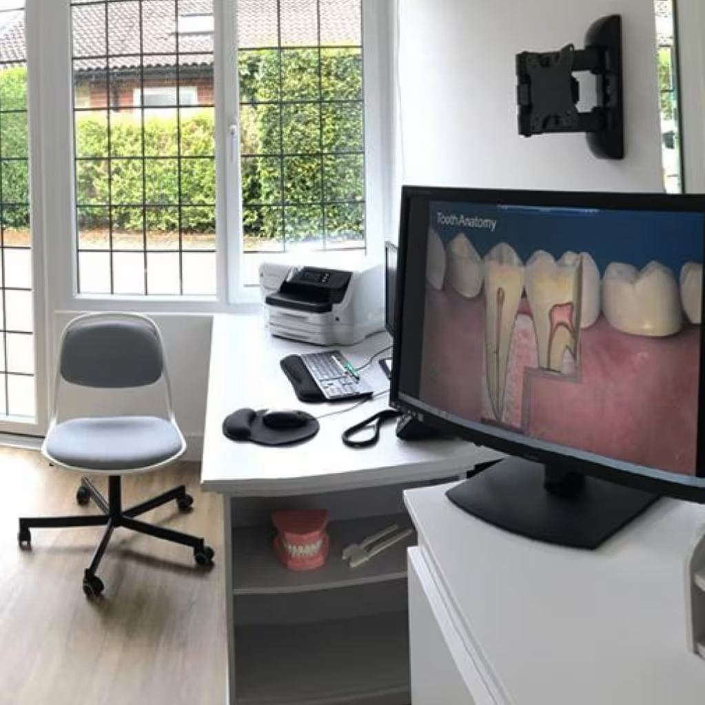
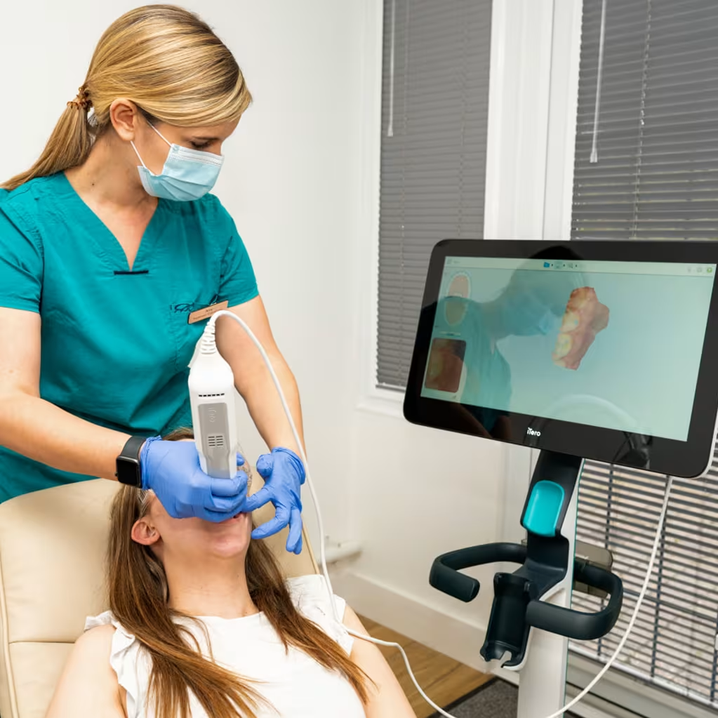
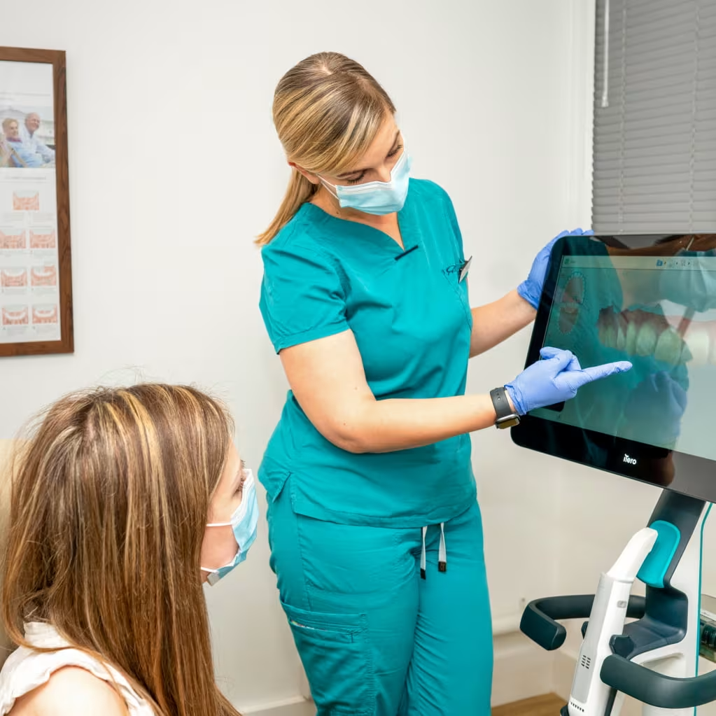
Dental x-rays serve as one of your dentist’s most powerful diagnostic tools, revealing critical information that can be completely invisible to the eye during a regular clinical examination. Without these images, dentists would be working blind to many oral health conditions developing beneath the surface.
Your mouth contains complex structures that simply cannot be assessed through visual inspection alone. X-rays penetrate through your teeth and gums to show bone levels, root structures, and developing problems that could become painful and expensive if left undetected.
Modern dental practices rely on these imaging techniques to provide preventative care rather than reactive treatment. This approach saves patients from unnecessary pain, complex procedures, and higher treatment costs down the road.
Bitewing x-rays are the most common type taken during routine dental visits. These images capture the crowns of your upper and lower back teeth simultaneously, providing crucial information about:
The name “bitewing” comes from the small tab patients bite down on to position the film or digital sensor correctly. These dental X-rays typically need updating every 12-24 months, depending on your individual risk factors.
Periapical dental x-rays show the entire tooth from crown to root tip, plus the surrounding bone structure. Dentists take these images when they need to investigate issues such as:
These dental X-rays provide the complete picture when specific teeth are causing problems or require detailed evaluation.
OPG x-rays capture a panoramic view of your entire mouth in a single image, showing all teeth, jawbones, and surrounding structures from ear to ear. This comprehensive imaging technique proves invaluable in several clinical situations:
Patient Comfort:
Diagnostic Applications:
Whilst OPG dental x-rays don’t provide the fine detail of intraoral images for detecting small cavities, they offer an excellent overview that guides further targeted imaging when necessary.
Clinical examination alone misses approximately 30-40% of cavities, particularly those developing between teeth. Dental X-rays reveal these hidden threats before they cause pain, infection, or require extensive treatment.
Cavities often start in areas completely invisible to the naked eye. The tight contacts between your teeth create perfect hiding spots for bacteria and acid production. By the time decay becomes visible clinically, it has often progressed significantly into the tooth structure.
Dental x-rays can detect these lesions when they’re still small and treatable with simple fillings, rather than requiring crowns or root canal therapy.
Periodontal disease destroys the bone supporting your teeth gradually and often painlessly. Dental x-rays show early bone level changes that indicate gum disease progression, allowing for timely intervention before tooth loss occurs.
Infections at tooth roots can develop without obvious symptoms initially. These periapical lesions, visible only on dental X-rays, can cause serious complications if left untreated, including facial swelling and systemic health issues.
Patient safety remains the top priority in dental radiography. Modern dental X-rays use extremely low radiation doses while providing exceptional diagnostic information.
The radiation exposure from dental X-rays is remarkably minimal in modern dentistry. For example, a complete set of bitewing X-rays delivers less radiation than a short commercial flight. For additional perspective, you receive more background radiation from natural sources in a single day than from a typical dental X-ray appointment!
Digital Technology Advantages
Digital dental X-rays have revolutionised patient safety by reducing radiation exposure by 80-90% compared to traditional film-based systems. This dramatic improvement means:
Cone Beam Computed Tomography (CBCT) represents the next level of dental imaging technology. While traditional dental X-rays provide two-dimensional images, CBCT scans create detailed three-dimensional views of your oral structures.
When Are CBCT Scans Necessary?
Dental professionals recommend CBCT imaging for specific advanced procedures:
These scans provide information impossible to obtain through conventional dental X-rays, enabling more predictable treatment outcomes and enhanced patient safety during complex procedures.
CBCT Safety Considerations
Whilst CBCT scans use higher radiation doses than traditional dental X-rays, they remain well within safe limits and are only recommended when the diagnostic benefits clearly outweigh the minimal risks.
Dental professionals follow strict guidelines about when dental X-rays are necessary, adhering to the ALARP principle (As Low As Reasonably Practicable) for radiation exposure, in line with UK radiation protection standards.
New Patient Evaluation
New patients typically require comprehensive X-rays to establish a baseline oral health status. This initial imaging reveals:
Risk-Based Scheduling
Your individual risk factors determine how frequently you need dental X-rays:
High-risk patients (every 6-12 months):
Low-risk patients (every 18-24 months):
Symptom Investigation
Dental X-rays become immediately necessary when you experience:
Dental X-rays remain an indispensable diagnostic tool that enables your dental team to provide comprehensive, preventive oral health care. These images reveal hidden problems that could otherwise develop into painful, expensive conditions requiring extensive treatment.
Modern digital dental X-rays combine exceptional diagnostic capability with minimal radiation exposure, making them safer than ever before. The radiation dose from dental X-rays is comparable to the natural background exposure you experience daily, with far less radiation than a commercial flight.
At The Briars Dental Centre, we follow strict guidelines about when dental x-rays are necessary, ensuring you receive appropriate care whilst minimising unnecessary exposure. This evidence-based approach protects your health whilst enabling early detection and treatment of oral health problems.
Trust your dental professional’s recommendation for dental X-rays—they’re an investment in your long-term oral health and overall well-being.
For more information, please contact our friendly team on 01635 40311 or reach out to us via email – enquiries@briarsdentalcentre.com. For further reading (without consulting Doctor Google), check out the British Dental Association’s information on dental radiography or PHE’s guidance!
Back to Blog